|
|
| Macrotrachela decora var. Bryce, 1912 |
|
| Macrotrachela decora, whirling. The diet of the specimens in this sample is desintegrating or reproducing cells of the green alga Trentepohlia (some of the cells can be seen below the rotifer in this image) |
| |
 |
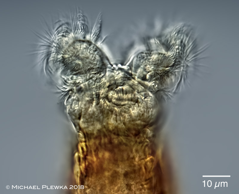
| Macrotrachela decora; head of whirling specimen; focus plane on the "minute fleshy ligules" (Bryce, 1912) in the sulcus (arrowhead). |
|
|
|
|
Macrotrachela decora var.; 4 aspects of the foot. The spurs are very short, cone-shaped with distinct tips (visible in the upper right image). The spurs seem to be appendages of a footplate. The top two images of the same specimen give the impression that there are two toes. On the other hand, the lower left picture shows a sheath which surrounds the two toes of the upper images together; a separation into two toes is not visible. This ambiguity of the findings is exactly what Donner (1965) describes for Macrotrachela decora. Lower right image: foot glands. |
| |
 |
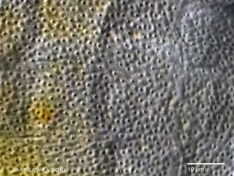
Macrotrachela decora; pores in the integument. |
| |
  |
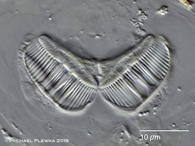
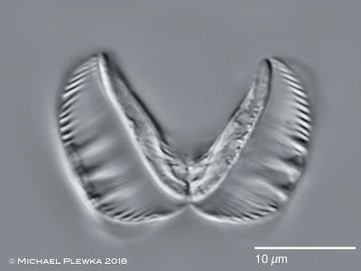
| Macrotrachela decora; two aspects of the ramate trophi. Left: cephalic view; dental formula 3/3; right: caudal view; ramus length (RaL): 16µm |
|
| |
| Description of BRYCE (1912):
"Specific Characters. -Of medium size and stoutness. Corona ample. Upper lip undivided, rising in a low curve, showing high nexus between divergent pedicels, widely separated. Rami with two to three teeth. Spurs short cones with moderate interspace.
Several examples of this moderately large form were obtained from moss growing on rocky outcrops near the top of Ben Vrachie, in Perthshire, in 1907. When creeping about they did not show any salient peculiarities, but in the feeding position the unusual form of the upper lip attracted attention. When feeding, the animals assumed a semi-squatting, rather trim and distinctive pose, in which the ample width of the corona became accentuated. The widely separated pedicels are distinctly divergent, although of moderate height. The trochal discs converge slightly to the median line, and have also a moderate dorsal inclination. The secondary wreath does not pass round the pedicels at their bases as is usual, but about one-third of their height up, a peculiarity which I think occurs also in Mniobia magna (Plate) and M. scarlatina (Ehr.). The nexus between the pedicels is exposed, and so high as to be nearly level with the inner margin of the trochal discs, Centrally it is decorated with two minute fleshy ligules. The upper lip rises in a very moderate curve nearly to the level of the nexus, and shows no trace of notch. As is not unusual, the post-oral segment is withdrawn within the following one when the animal is feeding, the dorsal antenna only being left visible. The rami appeared to be of normal form with the dental formula 2 / 2 + 1. A rather wide stomach lumen could be easily defined. The lumbar plicae persisted in the feeding position. The foot structure was not satisfactorily determined. My impression was that the toes were absent or modified to a sucker-like disc, in which case the species would properly belong to the genus Mniobia, and this relationship receives some support from the position of the secondary wreath. Pending further examination, it seems best to leave the species in the genus Callidina.
Maximum length not recorded; in feeding position, as figured, about 185 µ; corona, about 63 µ; collar, about 45 µ; spurs, about 5µ; interspace, 6 µ.Habitat, as stated above." |
| |
| |
| |
| Location: Arnsberger Wald; NRW, Germany; Hochmoor (Kapellenplatz) |
| Habitat: bark of fir tree, between Trentepohlia aurea |
| Date: 07.11.2018 |
|
|
|
|
|
|